Introduction
Introduction
Eight year old boy presented with lower limb edema, HTN, hematuria and proteinuria : 2.5g/day (normal :0.01-0.15g/day). A renal biopsy is performed.
Go to Question 1
Discussion
Discussion Case 17
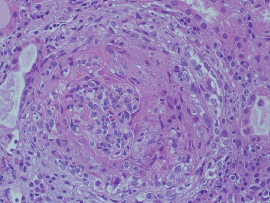
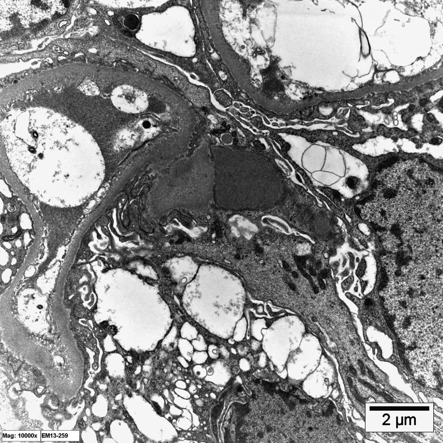
Question #1and 2: The first photomicrograph highlights the presence of extracapillary proliferation and in the center of the glomerulus, the compressed tuft is seen. By definition, the presence of this proliferation in more than 50% of the glomeruli qualify for a diagnosis of crescentic glomerulonephritis. Crescentic glomerulonephritis secondary to post-infectious glomerulonephritis is well documented and the presence in the second photomicrograph of a subepithelial hump support the diagnosis.