Introduction
This video demonstrates the tissue triaging procedure for renal needle core biopsies that are delivered already in a fixed/preserved state (10% buffered formalin/4% paraformaldehyde and Michel’s solution respectively.) Some labs may get tissue for EM in glutaraldehyde. The procedures demonstrated are: Specimen Identification (Universal Procedures as well as Procedures unique to Weill Cornell Medicine’s Renal Pathology Electron Microscopy Laboratory), Station Preparation and materials used, Initial Observation, Gross Tissue Examination, and Specimen Division for Histology, Immunoflourescence, and Electron Microscopy. In this demonstration, Paraformaldehyde in a Phosphate Buffer, pH 7.0 is used due to its unique ability to preserve tissue under adverse atmospheric and transport settings, sent from out of town areas. The fixed tissue from here can be used and divided for both light and electron microscopy in the laboratory. Preservative for fresh tissue is Michel’s solution for immunofluorescence. These solutions are available commercially and are provided to hospitals or nephrologists for uniformity of preservation and transport to our facility for processing. Other options are available and are at the discretion of the individual facility.
Video
Authors
 |
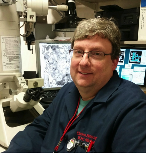
Michael Ganger Michael Ganger is the lead Electron Microscopist and EM Laboratory Manager at Weill Cornell Medicine for the past 15 years. Michael has a M.S. in Biology and is a Certified Biological Electron Microscopist with the Microscopy Society of America. He also is an Associate Professor at two local Universities: Montclair State University and New Jersey City University where he teaches Transmission and Scanning Electron Microscopy. |
 |
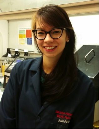
Sara Pawlak Sara joined the Weill Cornell Medicine EM team two years ago as an Electron Microscopy Technician. She has a B.S. in Environmental Sciences and is a Certified Biological Electron Microscopist by the Microscopy Society of America. Sara is actively pursuing her Masters Degree in Biomedical Laboratory Management with Hunter College in NYC. |
 |
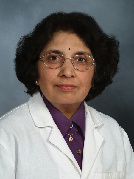
Surya V Seshan MBBS/MD Dr. Surya Seshan trained with Dr. Jacob Churg in Renal Pathology and is currently Professor of Clinical Pathology at Weill Cornell Medicine, New York, USA. She also serves as the chief of the Renal Pathology Division and Director of the Electron Microscopy laboratory at the Weill Cornell Medical Center/New York Presbyterian Hospital in New York. She is a past President of the Renal Pathology Society, a longtime member of the ISN and currently serving as a member of the ISN Pathology committee. |
Additional Info
-
Language:
English -
Contains Audio:
Yes -
Content Type:
Case/Images -
Source:
Other sources -
Year:
2015 -
Members Only:
No
